Cellular biologists from the University of California, Santa Cruz, have presented compelling evidence in a recent investigation, suggesting that an initial pregnancy occurring earlier in a woman’s life may confer protection against breast cancer for several decades thereafter. This protective effect appears to stem from the prevention of age-related cellular alterations within mammary tissues, which are known to be precursors to tumor development. By employing a murine model engineered to recapitulate human aging patterns and reproductive timelines, the research team observed that gestation profoundly influences the aging trajectory of mammary gland tissue. Specifically, it diminishes the accumulation of aberrant cells that possess the capacity to undergo identity changes, potentially initiating cancerous growth in later years.
This groundbreaking work, recently documented in the esteemed journal Nature Communications, seeks to elucidate a persistent enigma within the field of breast cancer biology. While established scientific understanding acknowledges that advanced age elevates the risk of breast cancer, it is also recognized that early-life pregnancy offers enduring protective benefits. The precise cellular mechanisms underlying this phenomenon have, until now, remained largely elusive.
Through the application of advanced single-cell analytical techniques, the researchers conducted a comparative analysis of mammary tissues from aged mice that had experienced pregnancy versus those that had not. Their findings revealed that mammary tissue, in the absence of pregnancy as it ages, tends to accumulate a specific population of cellular entities described as “confused” hybrid cells. These cells exhibit characteristics of attempting to simultaneously embody two distinct cell types and emit a pro-inflammatory signaling molecule known as IL-33.
The presence of IL-33 has the potential to instigate uncontrolled cellular proliferation, a foundational step in the genesis of tumors. Nevertheless, the study’s outcomes unequivocally demonstrated that pregnancy functions as a critical “cellular reset mechanism,” effectively inhibiting the proliferation and accumulation of these distinctive confused cells.
By mandating that these cells commit to a singular function and adhere to it, pregnancy effectively preserves the ‘lineage integrity’ of the tissue. This observation implies that the protective efficacy of pregnancy is derived from its inherent capacity to preclude the aggregation of these hybrid cells from the outset – an aspect that is now the central focus of our ongoing research endeavors.”
Shaheen Sikandar, assistant professor of molecular, cell, and developmental biology and corresponding author of the study
Simulating Decades of Risk in a Murine Model
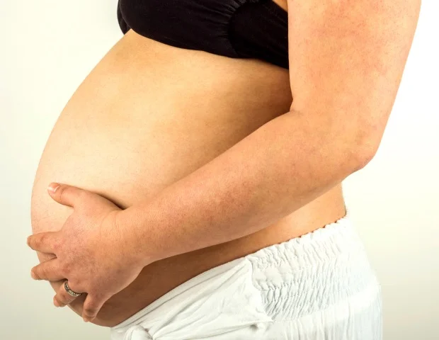
An individual’s susceptibility to developing breast cancer escalates consistently with age, with the majority of diagnoses occurring past the age of 50. Conversely, the parturition of a first child prior to reaching 30 years of age is associated with a diminished lifetime risk. To thoroughly investigate this correlation, the researchers’ investigation transcended the scope of most preceding studies, which primarily concentrated on the transient period immediately following pregnancy, a time when breast cancer risk is temporarily accentuated.
Instead, their examination delved into the state of mammary glands at a considerably later juncture, approximating postmenopausal age in human subjects. The experimental design involved a comparison between aged mice that had never undergone pregnancy (nulliparous) and aged mice that had experienced an early pregnancy. In terms of human analogy, this model is representative of women who gave birth to their first child between the approximate ages of 20 and 30, and whose mammary tissues were subsequently analyzed past the age of 50.
This extended temporal perspective is of significant consequence, given that approximately 75% of breast cancer diagnoses are made after the age of 50, according to comprehensive population-level cancer statistics cited by the researchers. Concurrently, the typical age range for a woman’s first pregnancy in the United States falls between 20 and 33 years.
Employing single-cell RNA sequencing technology, the research team meticulously analyzed thousands of individual mammary epithelial cells. This approach enabled them to track the intricate ways in which the combined effects of aging and pregnancy reshape cellular populations and modulate gene expression patterns.
A Perilous Cellular Subset Amasses with Age, Unless Pregnancy Intervenes
One of the most remarkable discoveries emerging from this study was the identification of these aforementioned hybrid cells. They are characterized as hybrid due to their co-expression of molecular markers distinctive to both the primary mammary lineages: luminal and basal. The anatomical positioning of these cells within the basal layer of the mammary gland further underscores their potential significance. A substantial body of existing research suggests that malignant breast tumors frequently originate from cells that progressively lose their normal cellular identity over time, a process particularly exacerbated by advancing age.
To ascertain whether the pro-inflammatory signaling molecule IL-33 itself could instigate detrimental cellular alterations, the researchers subjected mammary epithelial cells harvested from young mice to IL-33 treatment. The ensuing outcome mirrored the behavior observed in cells derived from aged, never-pregnant control animals.
Exposure to IL-33 resulted in heightened cellular proliferation and facilitated the formation of organoids – diminutive, simplified analogues of glandular tissue – particularly when this exposure was coupled with the suppression of Trp53, a critical gene functioning as a tumor suppressor. These observed functional modifications closely recapitulate key features associated with the initial stages of tumor development.
“Collectively, these findings offer a potential explanation for the protracted emergence of pregnancy’s protective effect, and why this protection endures well into later life. They achieve this by illuminating how early reproductive events can impart a lasting influence on the aging breast,” stated Andrew Olander, a graduate student within the Sikandar Lab and the principal author of this study.
Pregnancy Re-establishes Equilibrium and Fosters Cellular Differentiation
The influence of pregnancy extended beyond merely mitigating the quantity of hybrid cells. It also served to correct broader age-related imbalances present within the mammary tissue. In aged parous mice (those that had experienced pregnancy), the typical expansion of basal cells, a phenomenon commonly observed with aging, was normalized. Furthermore, both basal and luminal cell populations exhibited a diminished capacity to form organoids.
Simultaneously, luminal cells within aged parous mice retained molecular signatures indicative of “post-pregnancy involution,” a physiological state that may enhance their detectability by the immune system. The authors posited that this enhanced immune recognition could, in turn, bolster immune surveillance mechanisms and contribute to a further reduction in cancer risk.
Implications for Understanding Breast Cancer Risk Stratification
While this investigation was conducted using a murine model, the researchers assert that the underlying biological principles are highly likely to be pertinent to human physiology, given the established parallels in mammary gland architecture and breast cancer epidemiology. It is important to clarify that this study does not definitively establish a direct causal link between these hybrid cells and the onset of cancer. However, it undeniably identifies them as a probable contributor to age-related risk and a prospective target for the development of future preventative strategies.
“Our research provides a foundational understanding of the intricate interplay between aging and pregnancy within the mammary gland,” commented Sikandar. “Subsequent investigations will be dedicated to further elucidating the precise role of these ‘confused’ hybrid cells in the pathogenesis of breast cancer.”
Additional contributing authors from UC Santa Cruz include Paloma Medina, Veronica Haro Acosta, Sara Kaushik, and Matijs Dijkgraaf. Their research activities are situated within the Department of Molecular, Cell, & Developmental Biology, with some researchers also holding affiliations with the campus’s Genomics Institute, the Department of Biomolecular Engineering, and the Institute for the Biology of Stem Cells. The financial support for this research was generously provided by the Hellman Foundation, a National Institutes of Health/National Cancer Institute fellowship awarded to Olander, and a grant extended to Sikandar.
Olander, A., et al. (2026). Divergent aging of nulliparous and parous mammary glands reveals IL33+ hybrid epithelial cells. Nature Communications. DOI: 10.1038/s41467-026-68611-0. https://www.nature.com/articles/s41467-026-68611-0