Commonplace injuries known as splinters typically involve a minor fragment of material—be it wood, glass, metal, plastic, or a thorn—becoming lodged within the epidermis and the subcutaneous tissues.
The outermost layer of the skin, the epidermis, is rich in nociceptors. The dermis, situated directly beneath, possesses an even greater density of these sensory receptors, potentially rendering such penetrations exceedingly uncomfortable.
While the ability to extract a splinter may not be a critical survival skill, employing an effective technique can avert persistent discomfort or mitigate subsequent complications.
Scarcity in Medical Literature
Notwithstanding the paramount importance of pain management in healthcare, the phenomenon of splinters has garnered minimal scholarly attention.
In a 2004 publication, a collective of practitioners observed that “a paucity of controlled investigations exists comparing diverse removal methodologies, compelling physicians to depend on anecdotal accounts”. A contemporary examination of scholarly resources concerning splinters, conducted in 2025, yields predominantly a prolonged chronicle of individual case reports and subjective experiences.
Digital platforms and short-form video services are replete with “clever solutions” and recommendations advocating the use of substances like vinegar, adhesive tape, glues, onion segments, and banana peels, among other unconventional approaches. Evidence substantiating or disproving these practices is minimal, and some may indeed provoke cutaneous irritation or elicit adverse hypersensitivity reactions.
Ultimately, a sophisticated or unconventional remedy is not requisite for splinter eradication. The subsequent guidance outlines a methodical and secure approach to its removal, alongside criteria for seeking professional medical intervention.
Initial Assessment: Location Determined
The situs of the splinter serves as the primary determinant for the course of action. In instances where an ocular or palpebral splinter is suspected, immediate medical consultation is imperative, attainable via a primary care physician, an urgent care facility, or an emergency department.
Refrain from attempts to irrigate or flush the eye; this procedure necessitates execution by a trained healthcare provider using sterile saline solution within a controlled setting.
Fragments lodged beneath a fingernail or toenail, termed subungual splinters, frequently necessitate surgical intervention for their extraction.
Secondary Consideration: Composition Analysis
The material composition of the foreign body can also dictate the necessity for professional medical assistance.
Particular caution is warranted with glass fragments due to their propensity to fracture or splinter, leaving residual particles that prove challenging to dislodge and may precipitate persistent discomfort, inflammation, or infection.
External splinters originating from wood, thorns, or ferrous metals carry the potential risk of inducing tetanus, thereby necessitating a booster vaccination. Individuals with compromised immune systems or those who have undergone lymphatic surgery should seek prompt medical evaluation, as prophylactic antibiotic therapy may be indicated.
Essential Implements for Splinter Extraction
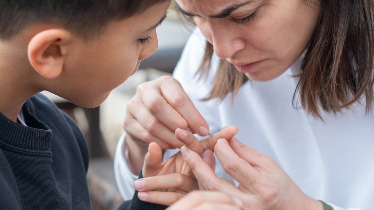
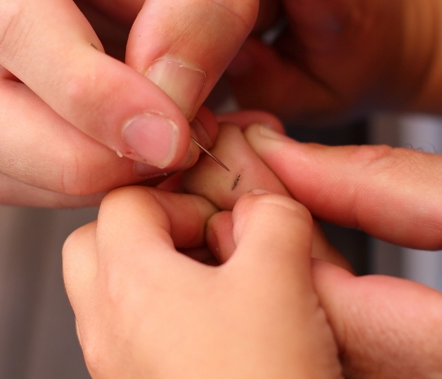
Should the aforementioned contraindications not apply, and the foreign body is clearly discernible, the most efficacious method of removal involves the use of fine-tipped tweezers.
If the extremity of the splinter is proximate to the epidermal surface, employing a beveled needle (available at pharmacies) can be beneficial to gently elevate the superficial skin layer, thereby exposing the embedded object. It is crucial to avoid penetrating deeper tissues, as this would inflict significant pain.
Prior to commencing the removal process, if the splinter is not composed of wood, immersing the affected locale in warm water may facilitate skin softening. While agents such as Epsom salts, baking soda, or hydrogen peroxide are sometimes recommended, empirical evidence substantiating their efficacy is lacking.
Avoid soaking wooden splinters, as this may induce swelling of the material, rendering its extraction more arduous.
Methodical Approach to Splinter Extraction
- Sanitize your hands thoroughly with soap and water or utilize an antimicrobial hand gel.
- Sterilize the tips of the tweezers (and needle, if employed) by cleansing them with the aforementioned sanitizing gel. Allow the instruments to air dry and avoid placing them on any surface prior to engagement.
- If visual acuity is a limiting factor, consider employing magnification aids such as reading glasses. This measure will prevent inadvertent contact with the splinter, thereby avoiding additional discomfort, and will facilitate a secure grasp with the tweezers. For metallic fragments exclusively, the use of nail clippers to gently grasp the splinter might enhance purchase.
- Extract the splinter by carefully retracing its path of entry—exert gentle traction in the opposite direction of its penetration.
- Upon successful removal of the splinter, cleanse the affected area with soap and water or an antiseptic solution. The application of alcohol-based hand sanitizer to the wound may induce a stinging sensation.
- In the event of superficial bleeding, apply an adhesive bandage or a small sterile dressing to the site.

For superficial splinters, visual confirmation of complete removal is generally achievable. However, for those embedded at a more oblique angle, assessing the totality of the extraction can be challenging. Deep-seated fragments may even necessitate radiographic imaging for precise localization.
Following splinter removal, vigilant observation for a period of several days is recommended to detect persistent discomfort or indications of infection, which may manifest as erythema, edema, increased pain, or purulent discharge. Untreated wound infections can escalate to sepsis, a critical and potentially life-threatening systemic condition.![]()