A remarkable digital reconstruction has brought to light the visage of a profoundly significant hominin fossil.
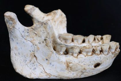
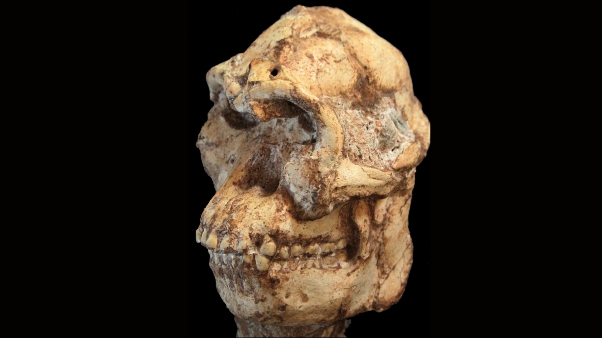
Referred to affectionately as ‘Little Foot‘, this 3.67-million-year-old specimen of Australopithecus presents an exceptionally complete skeletal structure. Its cranial vault, however, had suffered significant compression and distortion from millennia of entombment within a dense, rock-like matrix.
A groundbreaking investigation spearheaded by Amélie Beaudet, a paleoanthropologist at the Université de Poitiers in France, has successfully undertaken the first-ever digital reconstitution of Little Foot’s facial features.
Following this detailed reconstruction, the research cohort performed comparative analyses with other hominid and ape species. These examinations are instrumental in elucidating crucial aspects of our evolutionary facial development and that of our ancient progenitors.
The initial discovery of Little Foot dates back to 1980, unearthed within the Sterkfontein Cave system in South Africa. As its moniker implies, the earliest indications of its presence were four diminutive ankle bones. It was not until the 1990s that researchers identified the remainder of the skeleton embedded within the cave’s rocky strata, followed by a rigorous fifteen-year endeavor to meticulously extricate it from its fossilized encasement.
While generally classified within the Australopithecus genus, precisely assigning it to a specific species has proven challenging. This difficulty is partly attributable to the extensive deformation and fracturing of the skull, a consequence of geological pressures and movements within its stony confinement over geological epochs.
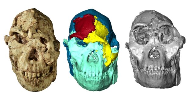
Consequently, the primary objective of this recent investigation was to restore the cranial structure to its pristine configuration. High-resolution X-ray micro-CT scans, conducted at the advanced Diamond Light Source synchrotron facility in the United Kingdom, yielded a three-dimensional digital rendering with an impressive resolution of 21 micrometers.
Subsequently, the skeletal elements and dental structures were virtually separated from the surrounding mineral matrix. The skull was meticulously divided into five distinct “blocks,” which were then manipulated within the 3D model, much like assembling a complex jigsaw puzzle, with the aim of reassembling them into their original topographical arrangement.
The research team proceeded to identify and meticulously measure key anatomical “landmarks” on the reassembled skull. This data was then subjected to shape analysis and juxtaposed with the cranial morphology of other Australopithecus individuals, as well as extant human skulls, and those of gorillas, chimpanzees, and orangutans.
Nonetheless, it appears to possess orbital regions with particularly distinctive configurations, potentially offering deeper insights into its evolutionary trajectory.
“It is plausible that evolutionary forces exerted specific selective pressures on the orbital region within southern African Pliocene hominins,” the scientists articulate. “This might have been associated with environmental instability, leading to diminished food availability and a greater reliance on fallback food sources that necessitated particular visual acuity.”
However, in keeping with the inherent complexities of research into the enigmatic hominin past, the investigators offer a note of caution regarding the definitive nature of their conclusions, citing several contributing factors.
The precise species attribution for Little Foot remains a subject of ongoing scientific discourse; it is even conceivable that it represents a previously undiscovered species. Furthermore, significant sexual dimorphism within the same species could potentially obscure accurate specimen classification.
The researchers acknowledge that their current reconstruction is “preliminary and would likely benefit from future refinements,” with certain distortional aspects proving intractable for complete correction. Such subsequent investigations hold the promise of bringing the physiognomies of our ancient kin into sharper relief.
This comprehensive research has been disseminated in the esteemed journal Comptes Rendus Palevol.