Utilizing artificial intelligence, investigators meticulously examined full-body MRI scans from an extensive cohort exceeding 66,000 individuals. This analytical endeavor culminated in the creation of the most comprehensive atlas to date, delineating the distribution of adipose tissue and musculature within the human physique, taking into account variations in age, biological sex, and stature. The findings of this research were disseminated today in the esteemed journal Radiology, an official publication of the Radiological Society of North America (RSNA). The study’s outcomes underscore that the integrity and quantity of skeletal muscle, in addition to visceral fat deposits, serve as potent indicators for the propensity towards developing diabetes, experiencing significant cardiovascular incidents, and overall mortality.
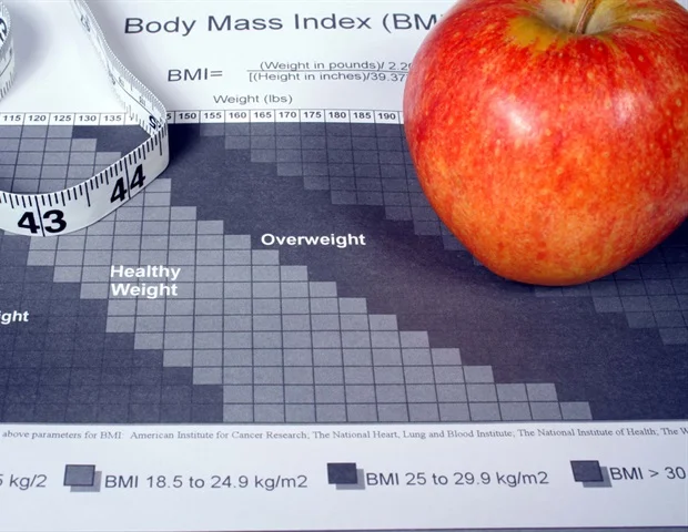
For a considerable period, medical practitioners have depended on metrics such as body mass index (BMI) and body weight to approximate the health risks associated with cardiometabolic disorders—the intricate interplay between the cardiovascular and metabolic systems—and general well-being. Nevertheless, BMI represents a rudimentary assessment of corporeal composition, deriving solely from height and weight measurements, and fails to incorporate the nuances of muscle mass or the spatial arrangement of adipose tissue.
Many risk assessment methodologies and therapeutic strategies continue to rely on BMI or waist circumference due to their ease of acquisition. However, BMI does not reliably represent an individual’s actual physical makeup.”
Jakob Weiss, M.D., Ph.D., senior author, radiologist in the Department of Diagnostic and Interventional Radiology, University Medical Center Freiburg, Germany
Dr. Weiss further elucidated that the medical fraternity also experiences a deficit in established benchmarks for how bodily composition evolves in individuals free from overt symptoms as they age, in addition to the disparities observed between males and females.
“There is mounting evidence suggesting that assessments of body composition function as independent risk determinants for cardiometabolic and oncological pathologies, as well as mortality,” remarked the study’s lead author, Matthias Jung, M.D., affiliated with the Department of Diagnostic and Interventional Radiology at the University Medical Center Freiburg. “Nonetheless, these metrics are influenced by an individual’s height and sex, and undergo substantial shifts with advancing age.”
The retrospective investigation encompassed a dataset comprising 66,608 participants (with an average age of 57.7 years, comprising 34,443 males, and an average BMI of 26.2). These individuals had undergone full-body MRI as contributors to the UK Biobank and the German National Cohort, with data collected between April 2014 and May 2022.
Leveraging their open-source, fully automated deep learning framework, the researchers derived body composition indicators from the MRI scans, meticulously normalized for age, sex, and height. The evaluated body composition parameters, encompassing subcutaneous adipose tissue, visceral adipose tissue, skeletal muscle, skeletal muscle fat fraction, and intramuscular adipose tissue, were quantified as z-scores. These scores denote the extent to which an individual’s measurement deviates from the established norm adjusted for age, sex, and height.
Subsequently, the research team performed rigorous statistical analyses to ascertain the prognostic significance of different z-score categories (low: z<-1; intermediate: z=-1 to 1; high: z>1) in forecasting the onset of diabetes, major adverse cardiovascular events, and all-cause mortality.
Their findings revealed a correlation between elevated visceral fat and a 2.26-fold amplified risk of future diabetes. Concurrently, increased intramuscular fat was linked to a 1.54-fold heightened probability of experiencing significant cardiovascular events in the future, and diminished skeletal muscle mass was associated with a 1.44-fold greater risk of all-cause mortality, independent of existing cardiometabolic risk factors.
“The critical factor is not solely the volume of muscle present, but also its qualitative aspect,” Dr. Jung emphasized. “Quantifying the amount of fat within the muscle provides insight into muscle quality that methodologies like BMI, bioelectrical impedance analysis, or DEXA cannot readily offer.”
The investigative team also formulated normative curves, adjusted for age, sex, and height, for key metrics of body composition.
“Accounting for extraneous variables is paramount for enhancing screening precision and tailoring therapeutic interventions,” stated Dr. Weiss. “This instrument possesses the capacity to identify instances where an individual’s body composition exposes them to a heightened susceptibility to metabolic disorders when contrasted with their age-matched contemporaries.”
In furtherance of scientific inquiry and to facilitate clinical application, the researchers have made their open-source, web-based calculator for age-, sex-, and height-adjusted body composition z-scores publicly available. This resource empowers researchers and clinicians to standardize their own datasets, thereby improving comparability and the generalizability of their findings.
“This tool enables clinicians to leverage routine imaging examinations opportunistically,” Dr. Weiss observed. “A dedicated full-body MRI may not be strictly necessary. If a standard CT or MRI scan of the torso already exists, the relevant data can be extracted for benchmarking against the established reference values.”
Dr. Weiss further posited that the AI-driven tool could contribute to refining risk stratification within oncology or differentiating between beneficial fat reduction and detrimental muscle depletion in patients undergoing treatment with weight-loss medications, such as GLP-1 agonists.
“We are already performing imaging on patients daily,” Dr. Weiss pointed out. “On every abdominal or thoracic scan, the requisite information is present, yet we do not routinely quantify or report it. Artificial intelligence now empowers us to access this latent data layer in a consistent and reproducible quantitative manner.”
The subsequent phases of the researchers’ work involve validating these normative curves within clinical patient groups, with a particular focus on predicting treatment toxicity, survival rates, and recurrence in cancer patients, as well as developing disease-specific reference values for additional patient cohorts.
Jung, M., et al. (2026). Body Composition in the General Population: Whole-body MRI–derived Reference Curves from Over 66 000 Individuals. Radiology. DOI: 10.1148/radiol.251939. https://pubs.rsna.org/doi/10.1148/radiol.251939