

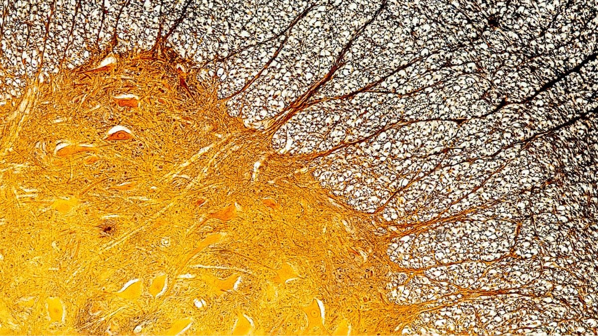
Receiving a diagnosis of multiple sclerosis (MS) often ushers in a period of considerable apprehension regarding the disease’s future trajectory.
Emerging scientific inquiry now illuminates a potential underlying mechanism and a therapeutic avenue for individuals experiencing the most severe manifestations of this condition.
MS is characterized by neuronal damage, which compromises the protective sheath encasing nerve cells, a crucial element for unimpeded signal transmission.
A recent investigation conducted by researchers in the Netherlands posits that in the gravest instances of MS, a specific type of immune cell, typically tasked with tissue regeneration and waste removal, becomes inundated with lipid accumulations.
These cells, identified as “foamy microglia,” have previously been observed in individuals afflicted with MS, although their precise role remained enigmatic.
The revelations from this latest study suggest that these lipid-laden microglia may indeed be principal contributors to the pathology of MS in its most advanced stages.
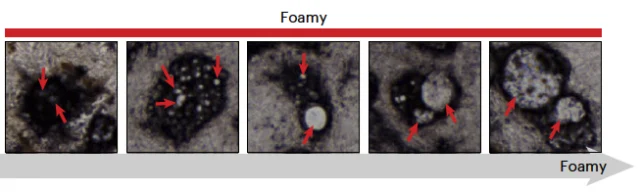
MS is classified as an autoimmune disorder where the body’s immune defenses erroneously target its own tissues, instigating inflammatory damage. However, the presence of these foamy microglia suggests a more complex etiology than inflammation alone.
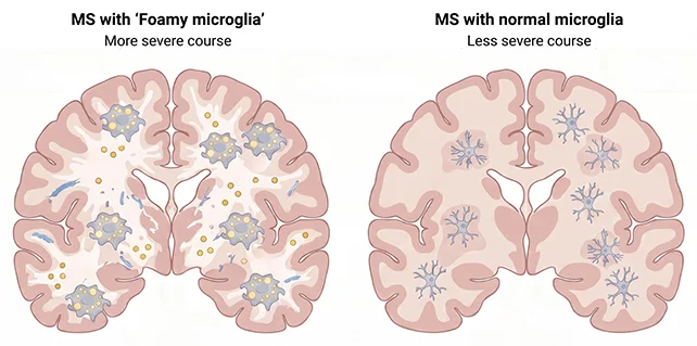
“We observed that patients with a significant abundance of these foamy microglia exhibited a more pronounced and frequent progression of the disease,” states Daan van der Vliet, a molecular physiologist affiliated with Leiden University in the Netherlands.
“It appears that the disease process is not solely attributable to the inflammatory response.”
The research team meticulously examined post-mortem brain tissue from 28 individuals diagnosed with secondary progressive MS, a phase characterized by a decline in cognitive and motor functions.
These samples were then juxtaposed with tissue from 10 donated brains of individuals without the disease.
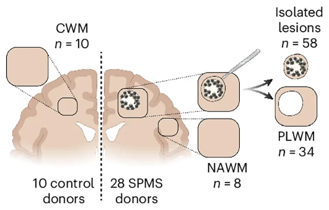
Employing a suite of sophisticated profiling methodologies, the researchers generated a comprehensive molecular atlas, delineating proteins, lipids, and gene expression patterns within the brain regions impacted by MS lesions.
These lesions arise when the myelin sheath, the fatty insulating layer surrounding nerve fibers, becomes the target of an overzealous immune response.
Beyond establishing a correlation between an increased presence of foamy microglia and MS progression, the study also revealed that these microglia were actively altering the inflammatory milieu surrounding the lesions, distinguished by a unique molecular signature comprising specific proteins and enzymes.
The researchers hypothesize that as microglia are dispatched to address neuronal damage, they become engorged with lipids, commencing with myelin, leading to cellular dysfunction and exacerbating the inflammatory cascade.
“It is probable that these cells are attempting a beneficial function: the clearance of damaged tissue,” explains van der Vliet.
“However, they become overwhelmed, so to speak. Consequently, their capacity to effectively contribute to repair is significantly diminished.”
Further bolstering these findings, the researchers utilized a murine model of MS and successfully inhibited a key enzyme highly active in foamy microglia. This intervention led to improved tissue regeneration in the mice, reinforcing the link between these immune cells and aggravated MS progression.
While these investigations are still in their nascent stages, subsequent clinical trials involving MS patients will be imperative to validate the observed association between foamy microglia and disease severity.
Further research will also be required to elucidate the long-term evolution of unrepaired lesions.
Nevertheless, these findings offer encouraging insights into the factors that differentiate individuals with MS who maintain a relatively normal quality of life for extended periods from those who experience premature paralysis or develop more debilitating symptoms at a younger age.
The research cohort expresses optimism that these discoveries could pave the way for novel therapeutic strategies targeting lipid metabolism within cells for MS management.
Additionally, this research, in conjunction with other ongoing studies, holds the potential for earlier identification of more severe MS cases.
The researchers identified traces of lipids associated with foamy microglia circulating within the cerebrospinal fluid, a finding they suggest could serve as a diagnostic biomarker for the disease.
“This opens the prospect of developing future biomarkers that could assist clinicians in discerning earlier which patients are at risk of rapid functional decline and, consequently, which therapeutic interventions would be most appropriate for them,” indicates van der Vliet.