

Not all areas of the brain exhibit an equivalent predisposition to oncogenic development.
Across numerous decades dedicated to the management of cerebral malignancies, medical practitioners have observed consistent tendencies—certain neoplasms invariably manifest within specific cerebral locales.
For instance, aggressive glioblastomas are frequently encountered within the cerebral hemispheres, whereas medulloblastomas typically originate in the cerebellum of pediatric patients.
For a considerable period, scientific inquiry has posited that specific neural tissues might inherently possess a greater susceptibility to cancerous transformation than others; however, the precise underlying mechanisms have remained elusive.
Presently, in the neural architecture of fruit flies, a research collective has unearthed a crucial revelation that could potentially pave the way for the development of novel therapeutic interventions for particularly virulent forms of brain cancer.
“Genetic alterations associated with oncogenesis occur routinely within our physiology, yet the vast majority do not escalate to a dangerous state because the organism’s defense systems identify and eliminate these aberrant cells,” observes oncologist Louise Cheng from the Peter MacCallum Cancer Center in Australia.
“Our primary objective was to elucidate the reasons why certain cells evade this surveillance and progress to form tumors, particularly within discrete brain regions.”
In contrast to many other bodily tissues, the living human brain presents considerable challenges for experimental investigation during the progression of disease. While meticulous clinical observation and the examination of donated neural tissue have provided substantial insights, much remains to be understood.
A remarkable parallel can be drawn from the unassuming fruit fly (Drosophila).
To probe the differential behavior of identical cancer-inducing mutations across various brain locations, researchers genetically engineered fruit flies. This manipulation targeted proteins instrumental in preserving neuronal identity, thereby inducing mature neurons to revert to a pluripotent, stem-cell-like state characterized by unchecked proliferation.
This established methodology for inducing tumorous growth in flies yielded rapid results, with the insects soon exhibiting burgeoning accumulations of abnormal neural cells.
However, an unexpected pattern subsequently became apparent.
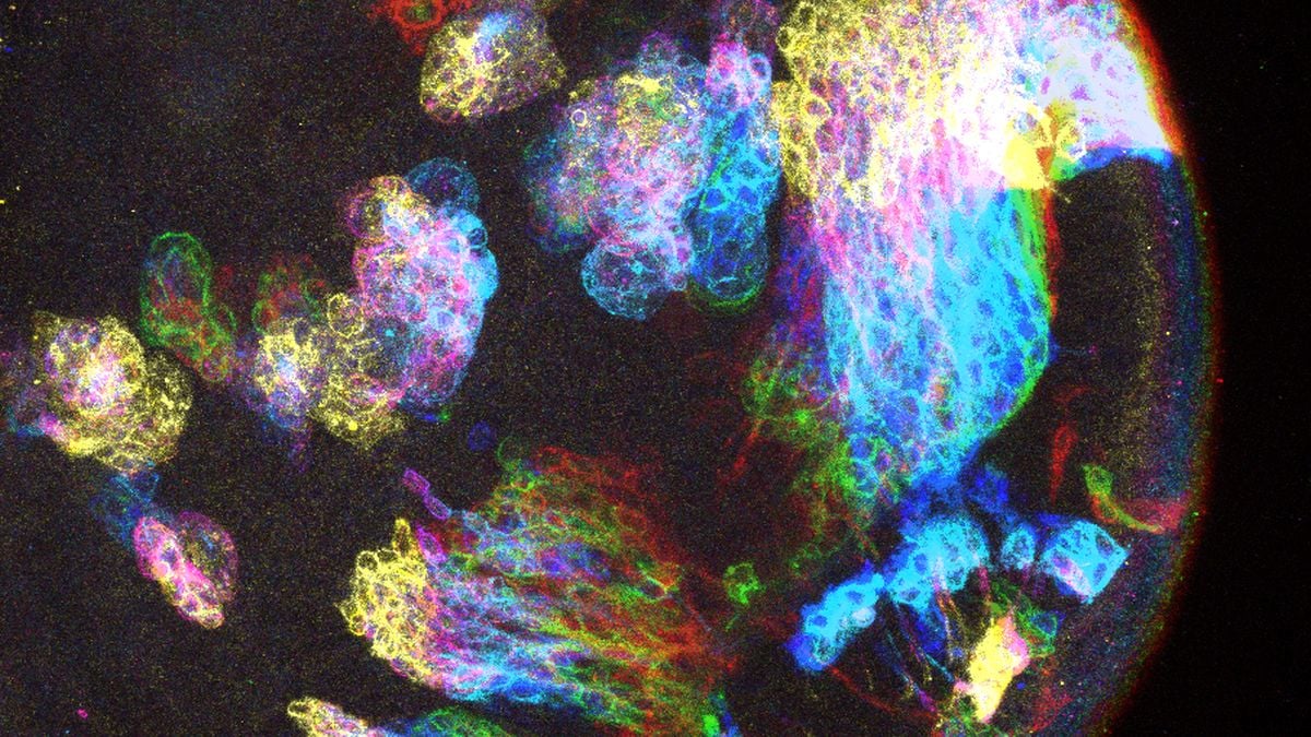
Although the undifferentiated, stem-like cells were distributed throughout the fly’s central nervous system, clinically significant tumors only persisted in select cerebral areas.
This observation strongly indicated a divergence between brain regions vulnerable to neoplastic development and those where such growth was inhibited.
Prior investigations had identified a specific protein, known as Chinmo, which plays a regulatory role in the maturation of progenitor cells.
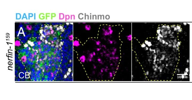
Upon quantifying Chinmo levels within the insect nervous system, the researchers detected a significant correlation.
In the central brain and ventral nerve cord, where tumors were observed to form, the aberrant cells exhibited Chinmo presence; conversely, in the optic lobes, which remained tumor-free, Chinmo was notably absent.
The subsequent phase involved the deliberate downregulation of Chinmo in regions predisposed to tumor formation and, in a separate experimental arm, its upregulation in the optic lobes.
The outcomes were remarkably pronounced.
In areas where Chinmo expression was suppressed, tumor progression was effectively arrested; conversely, where Chinmo levels were elevated, abnormal cell proliferation occurred in previously unaffected sites.
“We demonstrated the capacity to influence the cellular fate of genetically identical cells by modulating Chinmo activity,” Cheng states.
“Our findings underscore that tumorigenesis is not solely dictated by the mutation itself but is also substantially influenced by the cellular microenvironment and developmental stage at the site of mutation occurrence.”
While humans do not possess the Chinmo protein, rendering the findings an imperfect direct parallel, the study provides compelling evidence for discernible biological factors contributing to the higher incidence of tumors in certain brain areas.
Furthermore, it suggests the potential existence of analogous human proteins that exert similar influences on cancer susceptibility.
“Elucidating these determinants offers a novel framework for conceptualizing oncogenesis in humans beyond mere genetic mutations,” Cheng elaborates.
“By identifying the specific conditions that facilitate the transition of mutated cells into malignant tumors, we may unlock avenues for targeted therapeutic strategies to prevent cancer development.”