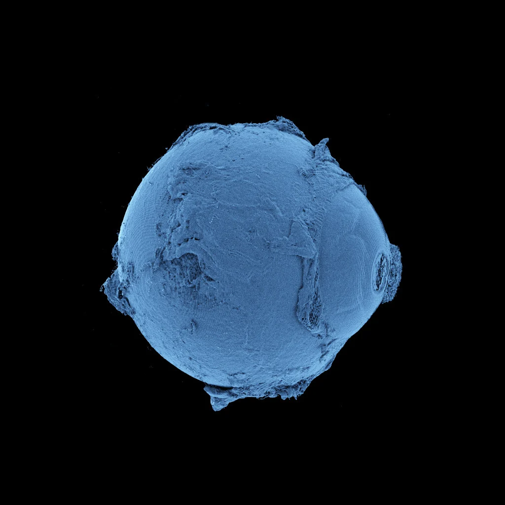
A groundbreaking initiative has provided an unparalleled view of the human physique, spanning from complete internal structures to minute cellular formations, with an exceptional degree of accuracy at a single micron resolution – a dimension approximately 50 times finer than a human hair.
Our biological systems can be conceptualized as intricate, layered constructs, comprising a hierarchical arrangement of cells, tissues, and organs. The very architecture of these components is fundamentally linked to their operational capacity, their interrelationships, and their susceptibility to pathological conditions.
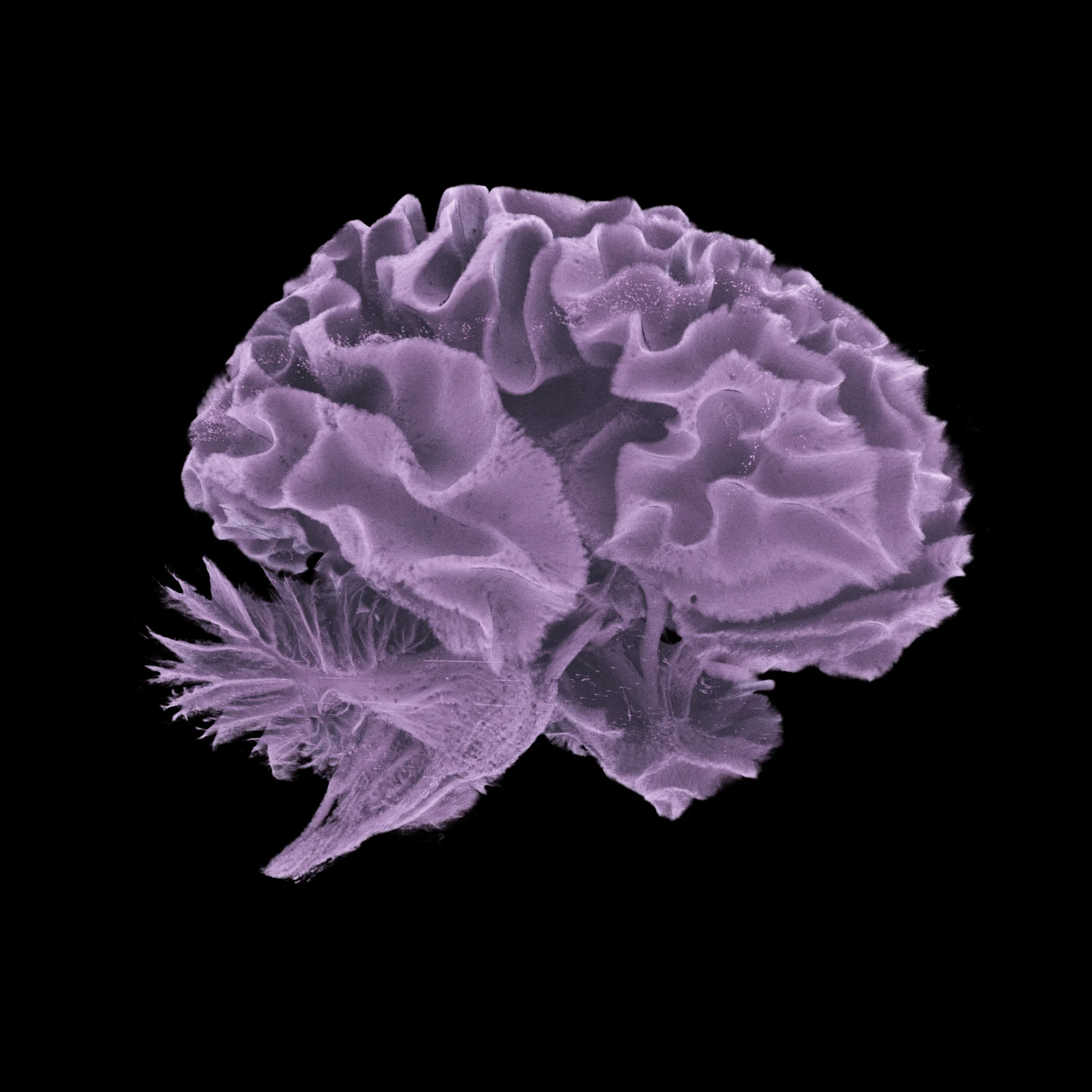
Currently, the Human Organ Atlas (HOA) is establishing a “new benchmark” in medical visualization by presenting the intricate frameworks of the brain, heart, lungs, liver, kidneys, and other physiological systems in exquisite, three-dimensional detail.
The HOA endeavors to make extensive scientific datasets, including numerous images exceeding one terabyte (TB), widely accessible. To provide context, a single TB is equivalent to over 250,000 photographs, 17,000 hours of audio recordings, or approximately 85 million Microsoft Word documents.
“This serves as a valuable repository for researchers, medical practitioners, and educators, as well as for any individual with a curiosity about the composition of the human body,” states Paul Tafforeau, a lead scientist at the European Synchrotron Radiation Facility (ESRF) in France, who spearheaded the advanced imaging methodology employed in the creation of the HOA.
The technique, known as hierarchical phase-contrast tomography (HiP-CT), utilizes X-rays generated by high-energy particles accelerated within a synchrotron, specifically the Extremely Brilliant Source (EBS).
This cutting-edge, fourth-generation particle accelerator provides a medical imaging capability that is up to 100 trillion times more potent than conventional hospital X-ray equipment.
To date, HOA researchers have employed the EBS to perform non-invasive imaging of intact, *ex vivo* organs from numerous donors, thereby achieving an exceptional level of magnification with cellular-level resolution.
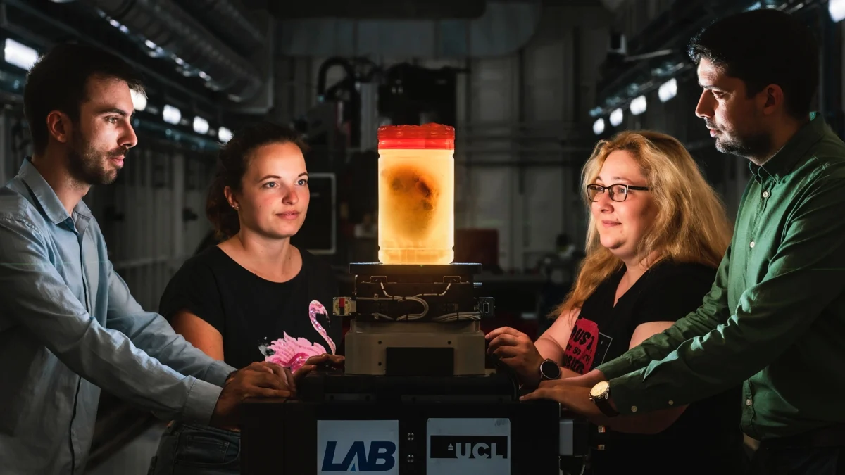
“The development of the Human Organ Atlas was a collaborative effort involving scientists and medical professionals from nine international institutions,” explains Peter Lee, a materials scientist at University College London (UCL).
“This consortium continues to grow, facilitating novel insights into conditions ranging from osteoarthritis to cardiovascular disease and fundamentally altering our approach to understanding human biology.”
Prior investigations employing HiP-CT imaging have uncovered previously unrecognized disease mechanisms at microscopic resolutions. These have included the identification of vascular damage in the lungs of individuals deceased from COVID-19, as well as the delineation of vascular characteristics associated with adenomyosis, a benign gynecological disorder.
As of the current date, the continuously evolving HOA features 87 distinct organs and 363 three-dimensional datasets, derived from the contributions of 54 donors.
In certain instances, the HOA incorporates imaging data from multiple organs sourced from a single individual. This is particularly valuable for analyzing the systemic impact of conditions like hypertension, as demonstrated by data from a donor with a history of high blood pressure, thereby enabling clinicians to study cross-organ effects – a key research objective.
A spectrum of other medical conditions is also documented, encompassing cancer, a significant cause of mortality in developed nations, as well as rare congenital anomalies such as Dandy-Walker syndrome, an infrequent condition affecting fewer than 1 in 30,000 live births.
Beyond its utility in medical education and training, the HOA possesses considerable potential for the development of machine learning algorithms, which are increasingly integrated, with varying implications, into contemporary healthcare practices.
Leveraging such a comprehensive, high-fidelity dataset for the training of artificial intelligence could lead to advancements in diagnostic accuracy and the refinement of therapeutic methodologies.
“I am immensely enthusiastic about the prospective applications of the Human Organ Atlas within the artificial intelligence community, particularly for the creation of foundational AI models,” expresses Claire Walsh, a biophysicist at UCL and director of the HOA Hub.
By illuminating the hitherto unexplored intricacies of human physiology, the research team anticipates that their work will foster greater public engagement with scientific endeavors, with further advancements anticipated in the near future.
“Currently, our focus is on individual organs. However, we foresee the evolution of this technology to enable the imaging of entire human bodies with a resolution ten to twenty times greater than current capabilities,” explains Tafforeau.
“Such data has the potential to revolutionize the study and comprehension of human anatomy.”