Scientists are leveraging medical imaging advancements, originally developed for live subjects, to meticulously unravel the enigmas of deceased individuals.
The examination of ancient Egyptian mummified remains, originating from bygone eras, was once a procedure that offered no assurance of preserving the cadavers in their entirety.
Presently, sophisticated imaging apparatus, akin to that found in hospitals, facilitates a comprehensive, layer-by-layer revelation of the contents concealed within ancient funerary wrappings, all while leaving the linen bandages undisturbed.
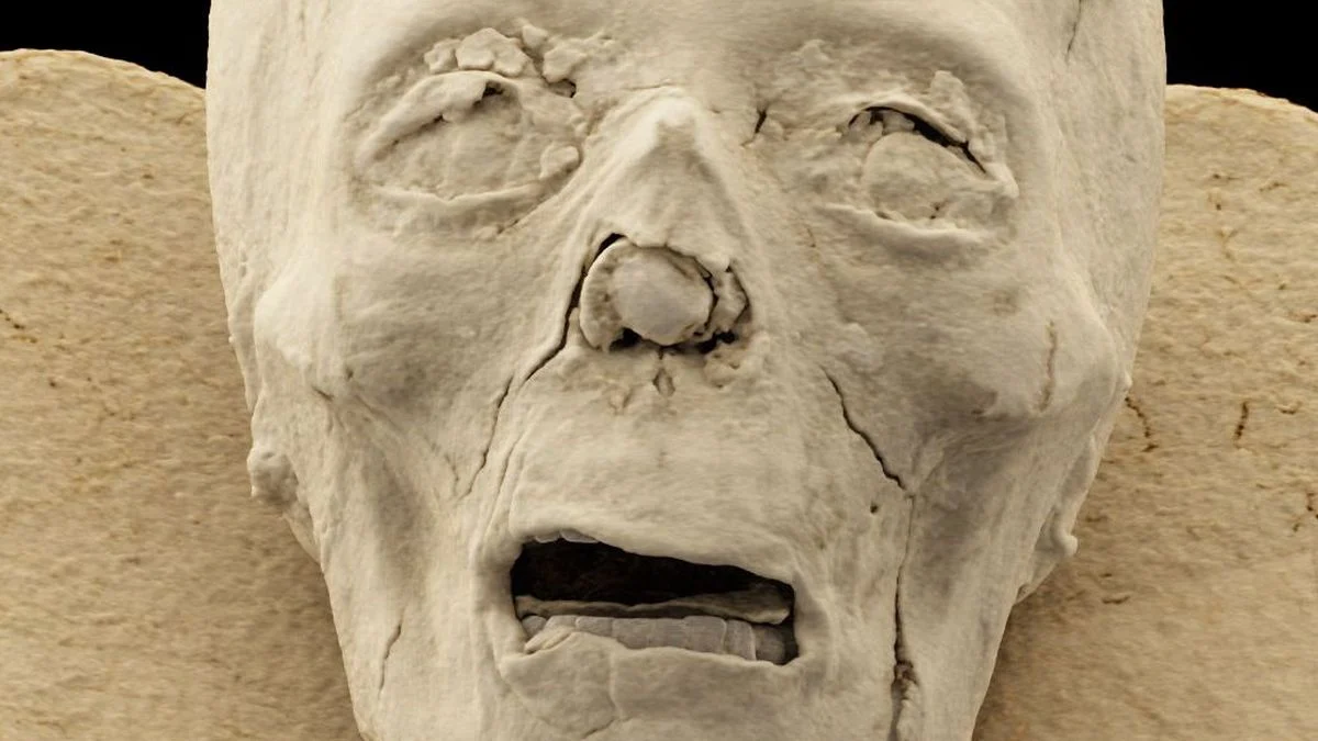
Upon the establishment of the Semmelweis Museum of Medical History, a branch of the Hungarian National Museum Public Collection Center in Budapest, in 1965, a collection of ancient Egyptian mummies was acquired. Researchers now aspire to decipher the profound secrets held within these specimens through the application of high-resolution computed tomography (CT) scanning.
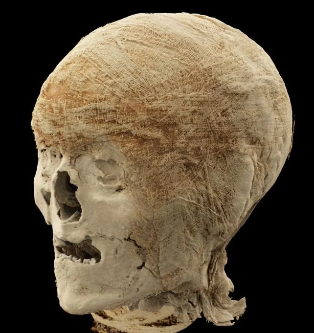
“The objective of these investigations is to generate the most precise possible depiction of the internal composition of the remains, identify any pathological conditions, and understand the preservation methodologies employed,” states Ibolyka Dudás, a chief clinical physician and radiologist affiliated with Semmelweis University’s Medical Imaging Center.
Computed tomography (CT) scanning technology has yielded remarkable benefits across numerous scientific disciplines. This advanced, high-resolution imaging technique utilizes rotating X-ray equipment in conjunction with computational analysis to produce detailed two- or three-dimensional visualizations of the internal structures of either inanimate objects or living organisms.
While the therapeutic applications of this technology are extensively documented and far-reaching, its utility has also been extended to scholarly pursuits, enabling non-destructive and non-invasive scrutiny of a diverse array of artifacts, including ancient paleontological specimens, intricate insect neural systems, and even extraterrestrial matter originating from the planet Mars.
Currently, Dudás and her scientific collaborators are employing the center’s latest CT scanner, outfitted with a cutting-edge photon counting detector, to augment understanding of the museum’s enigmatic mummified specimens.
“The artifacts had undergone previous evaluation by a research consortium,” explains Krisztina Scheffer, curator of the museum’s collection, “however, the contemporary imaging provides an unprecedented level of detail and is anticipated to yield novel, scientifically substantiated discoveries concerning the remains that have been housed within the collection for many years.”
Six of the mummies in the museum’s possession underwent carbon dating; however, only three yielded conclusive results. The oldest of these specimens dates back to between 401 and 259 BCE, though the practice of deliberate mummification of the deceased by Egyptians had been ongoing for several millennia prior to this period.
Although analyses of the scan data are currently in progress and have not yet been disseminated, other research initiatives employing CT scans to visualize similarly valuable ancient remnants have illuminated a great deal regarding the lifestyles of our ancestors.
Ancient Egyptian morticians frequently placed valuable objects, such as currency and talismans, within the layers of their funerary wrappings.
Radiographic examinations of other Egyptian mummies have brought to light various health afflictions, including degenerative joint disease, and one study documented a significant incidence of anemia among ancient Egyptian youth.
CT scans of antiquity’s remnants can also expose latent malignancies, and a singular CT investigation of pre-Columbian South American mummies served to reveal evidence of a violent massacre.
Preliminary examinations of the mummies at the Semmelweis Museum indicate that CT scans possess the potential to aid in the chronological classification of certain remains, based upon the embalming techniques that were employed.
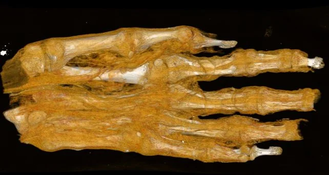
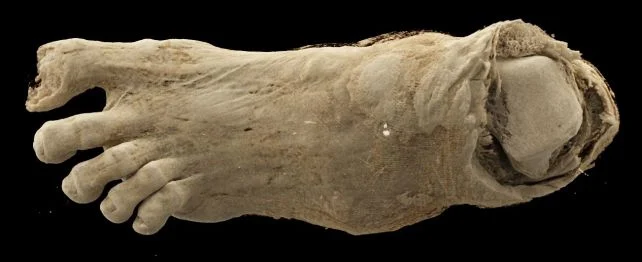
One particular specimen presented a noteworthy challenge – an unidentifiable bundle whose external appearance offered no clues to its contents. Initially, researchers speculated it might be an avian specimen, as ancient Egyptians frequently mummified birds. Another hypothesis suggested it could be a head, but the ensuing scans ultimately revealed it to be a foot.
A radiographic assessment of a distinct mummified foot also suggests that the individual experienced bone density loss. Researchers anticipate that subsequent scans will facilitate the extraction of further specifics regarding the individuals and the preservation methods utilized for their bodies.
“The extant findings convincingly demonstrate that contemporary imaging technologies are opening up novel avenues for research into mummified remains,” Scheffer asserts.
“This technology permits the retrieval of information concealed within millennia-old artifacts without causing them any degradation.”